Introducing ERGO, a multi-disciplinary research group.
Working to improve healthcare and treatments through research and innovation.
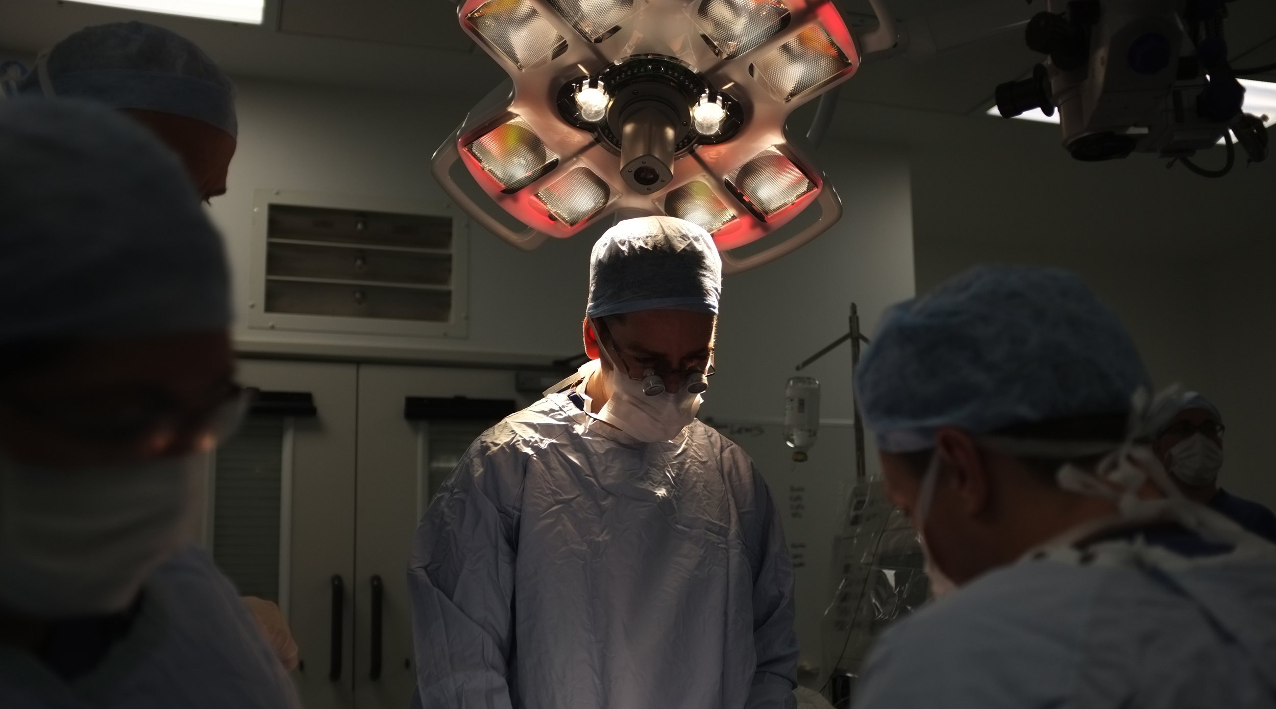
Our approach.
We are a multi-disciplined health care team supporting ophthalmology based research at the Oxford Eye Hospital. Our core goal is to contribute to advances in ophthalmology and related areas of medicine through innovative clinical research.
Clinical trials.
Our team of multi-disciplined health care professions support research trials from independent in-house projects to multi centre studies. We aim to deliver affordable solutions without compromising on the quality of care, keeping the patients needs at the forefront of our service.
You could help us with our research.
There are ongoing studies at the Oxford Eye Hospital which may be relevant to your eye condition, either now or in the future.
Meet the team.
In 2009 we set up the Eye Research Group Oxford (ERGO) with the goal of creating a formal structure for research within the Oxford Eye Hospital. We have expanded from a team of 2 dedicated researchers to a hub of a talented multidisciplinary team.
“Vision isn’t just about sight. It’s the ability to see beyond the here and now.”
— Tim Reddish OBE, Paralympic Athlete, Chair of BPA & Study Participant